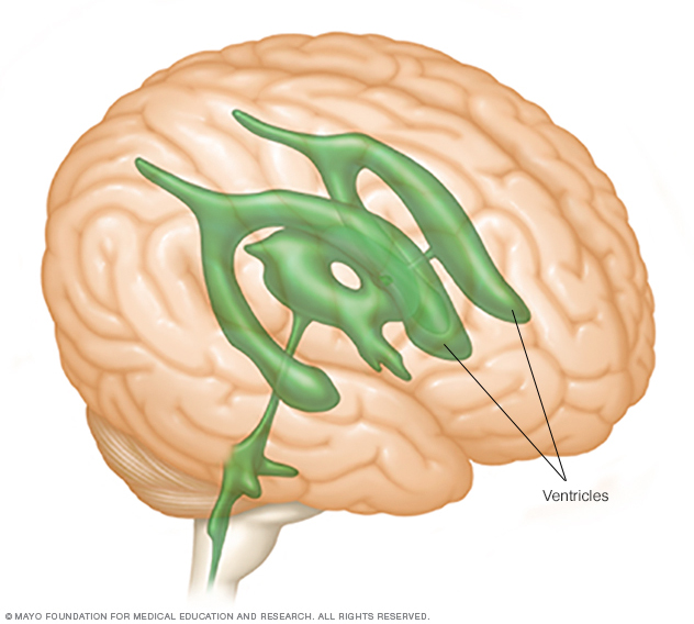

Hydrocephalus may also be present in a number of major and minor chromosomal aberrations affecting chromosome 8, 9, 13, 15, 18 or 21. The condition is characterized by aqueductal stenosis, severe intellectual disabilities and in half of the affected children, an adduction thumb deformity. Classic X-linked recessive hydrocephalus (Bickers-Adam syndrome) accounts for approximately 7 percent of male hydrocephalus. In its most severe form, the syndrome is often associated with other CNS, cardiac, genitourinary, ocular and facial anomalies and is often accompanied by intellectual disabilities. It is defined by hypoplasia of the cerebellar vermis, cystic dilatation of fourth ventricle and hydrocephalus. The Dandy-Walker malformation accounts for two to 10 percent of children with hydrocephalus. Affected children will have a variable level of paralysis of the legs and perhaps brain stem malfunction as well. This malformation is characterized by myelomeningocele and posterior fossa abnormalities, which have distinct sonographic appearances (the so-called “lemon” and “banana” signs). The Chiari II malformation accounts for approximately 30 percent of fetuses identified with ventriculomegaly. Blockage at the aqueduct is assumed when the lateral and third ventricles are enlarged proximal to the obstruction, and the fourth ventricle is relatively small.įetal hydrocephalus is also commonly associated with congenital malformation syndromes. It accounts for up to 20 percent of cases of fetal hydrocephalus. The most common form of isolated, obstructive hydrocephalus is so-called “aqueductal stenosis,” which is the blockage of CSF passage through the aqueduct of Sylvius. True fetal hydrocephalus has a variety of causes. Fetal ventriculomegaly is frequently associated with other severe developmental abnormalities, and this combination presents a uniformly dismal outcome. In these cases, such as hydrancephaly and holoprosencephaly, the ventricles are not only relatively enlarged but also often distorted due to overlying parenchymal abnormalities. The diagnostic distinction can be difficult to make, particularly with ultrasound alone, but is critical because fetal ventriculomegaly from a destructive or maldevelopment process carries a poor prognosis. It is important to distinguish hydrocephalus from ventricular enlargement or ventriculomegaly, which can also be caused by brain destruction and morphological maldevelopment. This knowledge has facilitated obstetric care but presents a source of uncertainty for families and a challenge for the team counseling parents regarding a prognosis for the fetus. However, with the advent of high quality prenatal ultrasonography, ventricular enlargement is now routinely diagnosed in utero. Traditionally, hydrocephalus is detected and treated after birth with a shunting procedure. Hydrocephalus is one of the most common congenital anomalies affecting the nervous system, occurring with an incidence of 0.3 to 2.5 per 1,000 live births. Ventricular shunting involves placement of a thin tube into the ventricles of the brain to drain fluid and relieve hydrocephalus. As the process continues, however, irreversible brain damage inevitably occurs. The brain can accommodate ventricular dilatation to a certain extent without significant neuronal damage. The ventricles begin to dilate, causing thinning and stretching of the cerebral mantle. If the CSF pathways are obstructed or obliterated by developmental or acquired abnormalities, CSF accumulates under pressure within the ventricular system.
#Ventricles of the brain series
By the neonatal period, approximately 300-500 cc of CSF is produced per day.ĬSF flows through a series of openings or foramens in the brain and out into the subarachnoid space where it is reabsorbed by the venous system. Within the ventricles, the choroids plexus produces cerebral spinal fluid (CSF) starting at the sixth week of gestation. As it grows, the inner tube remains and becomes a series of interconnected cavities known as ventricles.

During prenatal development, the brain starts out as a tubular structure.


 0 kommentar(er)
